
Eye Exam
INTRODUCTION
 The best way to ensure a lifetime of good vision is through regular, comprehensive eye examinations. In addition to ensuring proper eyesight, regular exams allow the early detection and treatment of diseases, improving the likelihood for successful treatment. Several eye diseases, such as glaucoma, show no symptoms in their beginning stages and can only be detected by a qualified examiner.
Not all eye examinations need be comprehensive. Some may be specifically formatted for the evaluation, diagnosis and treatment of a specific complaint. Generally, a comprehensive examination takes about hour, depending upon the number and type of tests required.
SOMEONE TO DRIVE YOU HOME
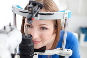
A comprehensive eye exam often requires that your pupils be dilated with eye drops. This enables us to thoroughly examine and accurately measure the eye. The drops that are used are longer lasting than what you may be accustomed to from past visits to the eye doctor.
These will leave your vision somewhat blurry and brighter than normal for about four hours. We recommend that you bring a pair of dark sunglasses and a friend or family member to drive you home afterwards.
SYMPTOMS AND COMPLAINTS
You will need to supply some basic information about why you are having an exam at this time. Here are some common questions you might need to answer:
- Why are you here?
- Did you bring your glasses or contacts?
- Do you have a specific problem?
- What are your symptoms?
- When does it happen?
- How often does it occur?
CASE HISTORY
Since the eyes are a part of the body, they can be affected by seemingly unrelated health conditions. You will need to supply information about your vision and health history, medications, symptoms of vision problems and other background information.
These are divided into 3 categories:
- Ocular: The physical condition of your eye, including past injuries, infections, surgeries, changes in vision correction.
- General: Lifestyle, including the past and current condition of your health, what medications you have taken, plus cigarette smoking, caffeine and alcohol consumption.
- Family: Family members with health issues such as high blood pressure, diabetes, thyroid dysfunction, coronary artery disease, or elevated cholesterol. These can be inherited and may show early signs in the eye.
EXAMINATIONS
- Visual Acuity (VA)
- External Examination
- Peripheral visual fields
- Pupil reflex tests
- Ocular motor tests
- Binocular vision tests
- Color vision tests
- Ophthalmoscopy
- Tonometry
- Keratometry
- Retinoscopy and Auto-refractometry
- The Subjective Refraction
- Phorometric (Phoria) Measurements
- Accommodative Range and Near Vision Analysis
VISUAL ACUITY (VA)
An assessment of the quality of your vision is called the Visual Acuity test, or VA. This test determines how well your eye makes images and is measured for distance and near vision with standardized eye charts, at standardized testing distances, under specific lighting. The Visual Acuity is noted as:- a fraction of the testing distance vs. the size of the letter
EXTERNAL EXAMINATION
This is an examination of the external parts of each eye and the surrounding associated tissues. Structures examined include: the eyelids, lashes, conjunctiva, iris, cornea, crystalline lens and anterior chamber structures. A slit-lamp biomicroscope is used for much of this exam. Gonioscopy, a detailed examination of the drainage system in the anterior chamber, may be included for people who may have glaucoma. This specialized test is generally not part of a routine examination.PERIPHERAL VISUAL FIELDS
This test may be performed by the use of a hand-held or by automated instrumentation, called the auto-perimeter test. It is included for people at risk for glaucoma or for routine “baseline” screening. Defects in the peripheral visual field may be an indication of internal eye conditions, including retinal detachment and glaucoma, or may be the result of problems with the brain or vascular system.PUPIL REFLEX TESTS
In this test, a light is shone into your eye and the iris is observed for its ability to constrict (reduce in size) and dilate (increase in size). Abnormal pupil reflexes may indicate faulty neurological (brain) development or could be a sign of an internal eye infection.OCULAR MOTOR TESTS
These tests measure how well your eye muscles work and whether they are working in harmony with each other.- In children, faulty ocular motor skills often leads to learning disabilities, as well as poor coordination with other motor skills such as walking or athletic abilities.
- In adults, if one is previously tested as normal and then suddenly not normal, this may indicate neurological disease, including multiple sclerosis, brain tumor, or result from stroke or other systemic diseases.
- Divergence is the ability of the eyes to turn outwards so that they are both aimed towards the viewed object.
- Convergence is the ability of the eyes to turn inwards so that they are both aimed towards the viewed object.
BINOCULAR VISION TESTS
This is testing to determine if both eyes are working together for accurate depth perception. “3-D” red/green or polarized lenses are often used for these tests.- In children, poor or lacking binocular vision may indicate overall faulty vision system development and lead to learning disabilities and “clumsiness”.
- In adults, if one is previously tested as normal and then suddenly not normal, this may indicate neurological disease, including multiple sclerosis, brain tumor, or the result of a stroke or other systemic disease.
COLOR VISION TESTS
The analysis of color perception is often tested on children but not necessarily on adults, unless there is reported change in color perception or a particular job requirement is involved (piloting for example). Changes in color perception for adults can be related to internal eye disease, neurological problems or drug toxicity.OPHTHALMOSCOPY
This is a test used for observing of the internal structures of the eye. It is the only technique aside from surgery that allows direct observation of the complete network of blood vessels. It can help diagnose hardening of the arteries, high blood pressure, diabetes melitis, as well as other whole-body and eye-specific disorders. It employs an instrument called an ophthalmoscope:- A direct ophthalmoscope is a handheld instrument brought very close to the eye. Using a light source to illuminate the inside of the eye, this instrument provides magnified but somewhat limited view of the inside of the eye.
- An indirect ophthalmoscope is a spectacle-mounted or headband mounted apparatus worn by the examiner, and is considered to be the standard of care. It is used to provide a significantly wider field of view in stereo (3-D). Drops are required to dilate (open up) the pupil. A large hand-held lens is held before your eye and Dr. Seibel (wearing the apparatus) is positioned a couple of feet in front of you. This allows him to examine the retina and optic nerve and the peripheral structures in the back of the eye, and the arteries and veins feeding them.A more recent variation of indirect ophthalmoscopy is performed with small hand-held lens at the slit-lamp. This method was pioneered by Dr. Seibel in the greater Los Angeles area and achieves superior magnified views to those of a direct ophthalmoscope.
TONOMETRY
This is the test for measuring the internal pressure of the eyeball. Elevated pressure is one of the signs of glaucoma. There are a number of instruments used to test eye pressure. Some require that an object be placed for a moment against the cornea in the front of the eye (usually requiring a drop of anesthetic and sometimes a liquid to color the tears) and some use a burst of air to press against the eyeball. Dr. Seibel utilizes instruments that do not use the air burst because they offer greater accuracy and comfort for his patients. Tonometry is often performed at the end of an examination.KERATOMETRY
This procedure measures the curvature of the cornea in two perpendicular meridians. The front of the eye may be round like a basketball, or irregular like a football (see Astigmatism). Retinoscopy and Auto-refractometry These are objective tests for test your capability to see images clearly, which don’t require your input and allow your examiner a good starting point for the determination of your lens prescription. Your subjective input is necessary in a separate test for refinement. A retinascope is a handheld instrument that allows the doctor to focus a spot of light onto the back of the eye through varying lenses. An auto-refractor does much the same thing automatically.THE SUBJECTIVE REFRACTION
This test requires your input. The examiner presents a series of lenses and asks for the your opinion as to “which is better, one or two?” There are no absolute answers; only whichever appears more visually clear to you at the time.PHOROMETRIC (PHORIA) MEASUREMENTS
The lenses that you choose during the subjective refraction are analyzed for their effects on eye muscle balance and coordination. You are asked to align a presented object or letter horizontally and then vertically. Sometimes, the lens that provides your best vision might cause your eyes to strain inwards or outwards and would not provide comfortable vision. Sometimes a slightly lesser power will be the better choice.ACCOMMODATIVE RANGE AND NEAR VISION ANALYSIS
Accommodation is the ability of the crystalline lens in your eye to flex and adjust its focus from distance to near, back and forth effortlessly. Your near vision is tested by using an eye chart, and a lens power that gives you clear and comfortable near vision is obtained. This may be the same as your distance lens power, or for those experiencing presbyopia it will be used for the bifocal or reading lens power. Scheduling an exam Aside from your routine exams there are several reasons to schedule a non-emergency visit. These include: gradual loss of ability to focus on near objects, blurred distance vision after concentrated near vision activities or the reverse, change in quality of vision with existing eyeglasses or contact lenses, headaches or eyestrain, especially after near vision tasks. For routine exams:- Infants and toddlers: by 6 months of age for a neurological, motor, gross vision evaluation. A knowledgeable pediatrician may be able to provide this service.
- Preschoolers: at ages 3 and 5 for a comprehensive vision exam.
- School age to 19 years: annual general screening for distance and near vision, sometimes provided by the school system. If normal, a comprehensive exam at age 7 or 8, pre-puberty, ages 14, 16 or 17 to be given by a qualified eye care professional.
- Adults: every 1 or 2 years – every 2 or 3 years if no lens correction is necessary; glaucoma test and eye health screening every year after age 35, or earlier if significant family history of eye problems.