About The Eye

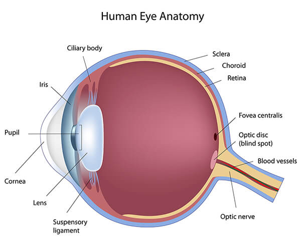
Being only about 1″ wide, 1″ deep and 0.9″ tall, the eye is a compact and amazingly complex organ.

The tough, outermost layer of the eye is called the sclera. This maintains the shape of the eye. The front part of this layer is clear and is called the cornea. All light must first pass through the cornea when it enters the eye. Attached to the sclera are six muscles that move the eye, called the extraocular muscles.

Chart showing the anatomy of the eye

  1. Conjunctiva
    Mucous membrane lining the inner surface of the eyelids and covering the front part of the sclera (white part of eye), responsible for keeping the eye moist.View Video
  2. Sclera
    White part of the eye. Tough covering that (with the cornea ) forms the external, protective coat of the eye.View Video
  3. Cornea
    Transparent tissue that forms the front part of the eyeball, covering the iris and pupil. The cornea is the first part of the eye that bends (or refract s) the light and provides most of the focusing power.View Video
  4. Iris
    Actually classified as a muscle, the iris is the colored part of the eye in front of the crystalline lens that regulates the amount of light that enters the eye by adjusting the size of the pupil in the center.View Video
  5. Pupil
    This is the black opening in the center of the eye where light enters. Pupil size changes when the iris tenses or relaxes depending on the amount of light present.View Video
  6. Crystalline Lens
    Double convex, transparent part of the eye, located behind the iris and in front of the vitreous body. Serves in conjunction with the cornea to refract incoming rays of light onto the retina.View Video
  7. Posterior capsule
    This is a thin membrane that holds the crystalline lens in place.
  8. Vitreous
    This is the gel that fills the eye and allows it to maintain its shape. It also serves as a clear pathway for light when it travels from the lens to the retina.
    View Video
  9. Macula
    Yellow spot on the retina, where the photoreceptors are most dense and responsible for the central vision. Has the greatest concentration of cones, responsible for visual acuity and the ability to see in color.View Video
  10. Retina
    The retina is the nerve center of the eye where light is converted into an electrical signal that travels to the brain. Cells, called rods and cones, within the retina transmit these signals along the optic nerve, thereby enabling sight.View Video
  11. Optic Nerve
    Bundle of nerve fibers that connect the retina with the brain. The optic nerve carries signals of light to the area of the brain called the visual cortex, which assembles the signals into images called vision.
    View Video

Seibel Vision Surgery provides this on-line information for educational and communication purposes only and it should not be construed as personal medical advice. Our mission is to provide an overview of these topics. For more in-depth information, please discuss any vision issues with Dr. Seibel or other eye care professional.

Information published on this web site is not intended to replace, supplant, or augment a consultation with an eye care professional regarding the your own medical care. Seibel Vision Surgery disclaims any and all liability for injury or other damages that could result from use of the information obtained from this site. Please read our full disclaimer.

Cataract Lens Icon

CATARACT SURGERY
advanced LENS IMPLANTS

Learn about cataracts and their treatment

Learn more about Cataracts
Crosshairs Icon

Laser Vision
Correction

LASIK, PRK, Implants, and other

Learn more about LASIK
Lightbulb Icon

OTHER EYE
CONDITIONS

Glaucoma, Macular Degeneration,
Medical Eye Exams, and other

LEarn More about Eye Conditions