As part of your LASIK preparation, you will need to receive a pre-operative eye exam. If you have any questions about why a particular test is included, please feel comfortable discussing it with Dr. Seibel until you are satisfied with the explanation. Your pre-operative LASIK exam will:
Address existing eye health concerns
Identify your potential surgical outcome
Frame expectations for what LASIK can do
Clarify your overall vision-health status
The pre-operative LASIK eye exam covers:
Assessment of your eye health history
Current prescription
Contact lens wear
Ocular or systemic diseases and medications
Previous ocular problems
Previous eye injuries
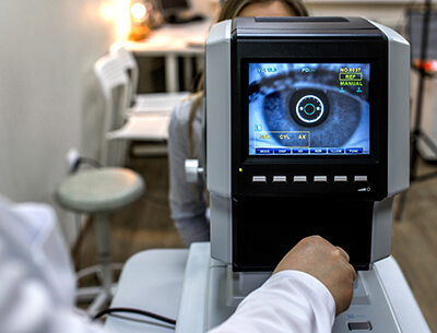
Pre-operative LASIK eye exam
Refraction (including dilation)
Motility
Eyelids
Conjunctiva
Cornea
Crystalline lens
Intraocular pressure
Posterior segment
Tear film
Pupil size
Epithelium
Corneal thickness
Topography
Wavefront LASIK
Lifestyle
Reviewing the results
If you are having your LASIK candidacy screening done through a your own eye care professional, you are still required to complete a pre-operative LASIK exam at our office before your LASIK surgery date. We will be unable to confirm your candidacy for LASIK or PRK until these tests are completed.
Someone to drive you home
A comprehensive LASIK eye exam requires that your pupils be dilated with eye drops. This enables us to thoroughly examine and accurately measure the eye. The drops that are used are longer lasting than what you may be accustomed to from past visits to the eye doctor.
These will leave your vision somewhat blurry and brighter than normal for about four hours. We recommend that you bring a pair of dark sunglasses and a friend or family member to drive you home afterwards. We also offer a type of eye drop that reverses dilation (reducing the time to about one hour), although many patients prefer not to use it because it usually causes side effects of irritation and redness.
Assessment of your eye health history
Current prescription: Your current prescription needs to have been stable during the last 2 years in order for LASIK to be appropriate at this time.
Contact lens wear: Contact lenses warp the corneal surface and lead to measurements that are not representative of your true refraction. You will have to stop wearing contact lenses prior to LASIK eye surgery. The time interval required depends upon the type of lens and how long you have been wearing them, and may be longer than the minimum times listed below depending on your initial topography results.
Soft lenses At least 2 weeks prior to your LASIK exam At least 2 weeks prior to LASIK surgery
Gas permeable and true hard (PMMA) lenses At least 3 weeks prior to your LASIK exam – if you have worn them for more than 10 years an additional week for each decade of wear is usually required. At least 3 weeks prior to laser eye surgery
Follow this advice in order to avoid unnecessary risk of complications or reduce the effectiveness of your LASIK surgery.
Ocular or systemic diseases and medications: Some ocular or systemic diseases and medications can affect your suitability as a candidate for LASIK.
Previous ocular problems: This refers to such conditions as lazy eyes, strabismus, or double vision and will need to be addressed in order to validate your candidacy for LASIK.
Previous eye injuries: Be sure to inform Dr. Seibel about any eye related injury, regardless of how long ago it occurred.
Your pre-operative eye exam:
Refraction: This test determines the visual acuity of your eyes, both uncorrected and as corrected by glasses or contacts, both with and without dilation.
Motility: This exam will assess the ability of the muscles to align your eyes properly.
Eyelids: This test exams your eyelids to see if they turn inward (possibly scratching the cornea) or outward (possibly directing tear flow away from the eye), plus other related conditions.
Conjunctiva: The conjunctiva is the transparent membrane that covers the outer surface of the eye and lines the inner surface of the eyelids. This test will check for irritations, redness, irregular blood vessels or other abnormalities.
Cornea: The cornea will be examined for overall health, scarring, any previous trauma, and pathology, using slit lamp via a microscope.
Crystalline lens: The natural lens inside your eyes will be examined to determine if clouding of the lens (a cataract) or other abnormalities are present.
Intraocular pressure: The intraocular pressure inside your eye will be measured to detect glaucoma or pre- glaucomatous conditions. Glaucoma is a visual loss caused by damage to the optic nerve from excessively high pressures in the eye. It is one of the most common causes of preventable vision loss.
Posterior segment: The dilated (fully open) pupil exam is used to assess the health of the posterior segment (inside back surface of your eye). This test allows Dr. Seibel to screen the retina, optic nerve, and blood vessels for a number of eye and systemic disorders.
Tear film: It is normal for patients to experience temporary dryness after LASIK. However, patients who have inadequate tears before surgery are at higher risk of prolonged dry-eye symptoms after surgery. Keep in mind that feeling of dryness while wearing contact lenses is not the same thing. Experiencing dry eyes while wearing contact lenses does not mean that the eyes are dry when contact lenses are not being worn. Different methods to evaluate tear function include:
Placement of colored agents in the tears
Physically measuring the tears themselves
Inspection of the tear film with a microscope
Pupil size: Most glare, halos, and diminished night-vision following LASIK surgery is due to a combination of larger pupil size and a high amount of nearsightedness or farsightedness. Because large pupils allow light rays to enter from the periphery of the cornea, custom programming the laser for a larger optical and ablation zone can minimize glare and halos. However, this can only be accomplished with lasers that have an adjustable zone from 6.0 mm to 8.0 mm, such as the ones we use here at Seibel Vision Surgery.Pupils should be measured when they are the largest potential size, which occurs under the darkest conditions. There are four methods for pupil size evaluation:
Light amplification – utilizing night vision technology derived from military devices, an examiner looks through one end of the device at the pupil. Pupil diameter is measured against a tiny ruler in the viewfinder to give accurate measurements in room light that would otherwise be too dark to take a measurement.
Ruler card – an examiner uses a hand-held card with different sized circles on it to match the diameter o the pupil to that of a circle on the card. This has the disadvantage of requiring that the room lights be bright enough for the examiner to read the card; therefore, the pupils may be smaller with this level of illumination than they would really be with nighttime illumination.
Visual estimation – an examiner guesses the pupil size by simply looking at it. This is the least accurate method and has the same disadvantage of the ruler card of requiring bright enough lighting for the examiner to see the pupils.
Epithelium: The cornea is covered on the surface with a thin clear layer of skin cells, called the epithelium. In some people, this layer of cells is not attached firmly. Loose epithelium may lead to a higher risk of complications. By microscopic examination of the cornea, this test will determine if a loose epithelium condition exists.
Corneal thickness: The medical term for measuring corneal thickness is pachymetry. This test will be used to calculate precisely how deep the laser will be allowed to penetrate. Since LASIK involves creating a flap on the surface of the cornea and using a laser to reshape the cornea by removing tissue, it is possible for the laser to remove too much, leading to corneal bulging and distorted vision. In order to be sure the laser does not penetrate beyond the safest level, pachymetry is imperative.
Topography: Using a Zeiss Humphrey Atlas 995 topographer, your eyes will be tested by corneal topography. As no one has a perfectly rounded cornea, the topographer machine will display the degree of astigmatism you have – the irregularities and actual steep or flat shape of your cornea, which cause your vision to be less than perfect. Occasionally patients have abnormal astigmatism called keratoconus, which is a cone-shaped cornea. These corneas often are weaker than normal corneas and thus should not be treated by LASIK.
Wavefront Mapping: The VISX CustomVue Wavefront Analyzer will provide a detailed analysis of your entire visual system. Dr. Seibel will use this information to further validate that you are an appropriate laser eye surgery candidate and formulate a highly customized vision correction plan specifically for your eyes. Read our section on Wavefront LASIK for more information on this incredible technology.
Vocational and lifestyle: Your work or recreational requirements can influence vision correction choices. For example, some choices can affect depth perception or the ability to see well at a particular distance.
Reviewing the results: You and Dr. Seibel will go over the results of your exam together and further plan your LASIK eye surgery.
Next step: Schedule and prepare for your LASIK procedure. This appointment will be scheduled at the time of your comprehensive eye exam.
At Seibel Vision Surgery, your eyes and vision are of paramount importance. To help you make the most informed decisions regarding the welfare of your vision, we are delighted to be of service and are happy to answer any questions you may have.
Please take the time to familiarize yourself with the information contained on this site. We have compiled it especially for you!
Monday, Tuesday, Wednesday, and Thursday 8:30 a.m. to 3:30 p.m. Friday 8:30 a.m. to 3:00 p.m.
OUR ADDRESS 11620 Wilshire Boulevard, Suite 711 Los Angeles, California 90025